Expert Perspectives
Dr. Bojun DONG
Department of Urology, Renji Hospital Shanghai Jiao Tong University School of Medicine
1Operative Protocol and Electrode Placement Strategy
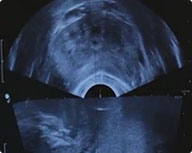
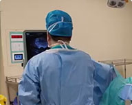
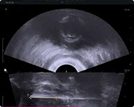
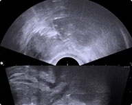
(1)Following general anesthesia, a transrectal ultrasound (TRUS) probe is inserted. The prostate is visualized in both axial and sagittal planes to assess its position and dimensions, with systematic image capture performed at 5-mm intervals.
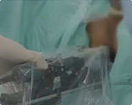
(2)Electrode selection is based on prostate dimensions, with ultrasound-guided insertion maintaining an inter-electrode spacing of 0.5-2 cm.
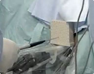
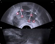
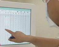
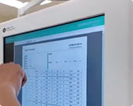
(3)The measured inter-electrode spacing is recorded and input into the system, which automatically generates ablation parameters for treatment execution.
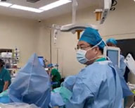
(4)Upon completion of voltage ramping, depress the footswitch to deliver pulses to the target lesion while monitoring real-time current output.
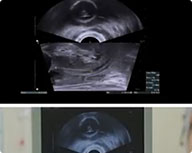
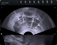
(5)Each pulse sequence requires approximately 5 minutes. Upon completion, the system automatically generates a treatment log, followed by post-procedural image acquisition at 5-mm intervals.
(6)The electrodes are withdrawn, followed by 1-minute compression hemostasis. After urethral irrigation with povidone-iodine solution, a urinary catheter is inserted and secured.